Diane Jacobs
Posted - 2007
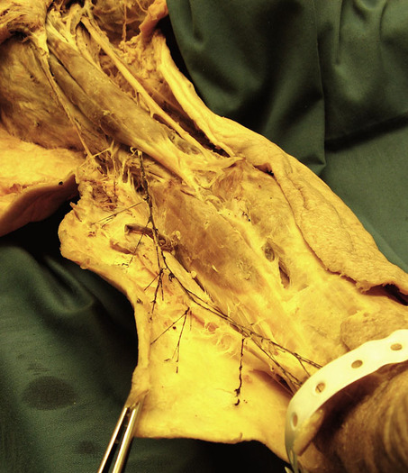
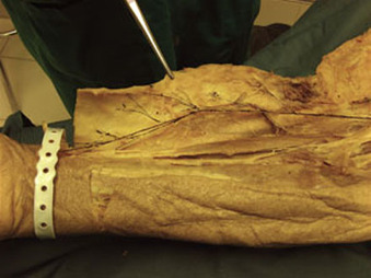
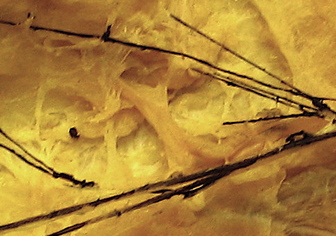
Cadaver dissection - musculocutaneous nerve. Diane Jacobs, PT, in British Columbia, Canada, has just released these hot pictures of her recent dissections of the cutaneous branches of the musculocutaneous nerve. The branches are colored black and can be seen to terminate in a disseminated fashion throughout the skin over the radial aspect of the forearm. The nerve leaves the lateral cord of the brachial plexus (C5, 6, 7) and innervates the coracobrachialis, biceps and brachialis muscles and the skin over the radial aspect of the forearm. During the median neurodynamic test, it becomes tight, where it can sometimes be seen as it passes across the lateral aspect of the elbow into the forearm. The cutaneous division can often be readily palpated next to brachioradialis and is sometimes mistaken for the posterior interosseous nerve (PIN), even though the PIN is deep to these structures. Diane is a spearhead of the formation of the fast-growing Canadian Physiotherapy Pain Sciences Group and has a particular interest in the relationships between skin innvervation and clinical pain states. We are grateful for the opportunity to present these illustrations and thank her very much.
Posted - 2007
Cadaver dissection - musculocutaneous nerve. Diane Jacobs, PT, in British Columbia, Canada, has just released these hot pictures of her recent dissections of the cutaneous branches of the musculocutaneous nerve. The branches are colored black and can be seen to terminate in a disseminated fashion throughout the skin over the radial aspect of the forearm. The nerve leaves the lateral cord of the brachial plexus (C5, 6, 7) and innervates the coracobrachialis, biceps and brachialis muscles and the skin over the radial aspect of the forearm. During the median neurodynamic test, it becomes tight, where it can sometimes be seen as it passes across the lateral aspect of the elbow into the forearm. The cutaneous division can often be readily palpated next to brachioradialis and is sometimes mistaken for the posterior interosseous nerve (PIN), even though the PIN is deep to these structures. Diane is a spearhead of the formation of the fast-growing Canadian Physiotherapy Pain Sciences Group and has a particular interest in the relationships between skin innvervation and clinical pain states. We are grateful for the opportunity to present these illustrations and thank her very much.