
New Model for Investigating Effects of Neural Techniques On Muscle Function During Manual Therapy
Rade, Shacklock, Peharec, Bačic, Candian, Kankaanpää, Airaksinen 2012. Journal of Electromyography and Kinesiology 22 (2012) 363–369
|
|
This article features interactions that we have found to occur between nerves and muscles during simulated Mills manipulation. The reason we feature this is that it is a new model for investigating interactions between neural and musculoskeletal tissues, providing new implications for clinical neurodynamics.
|
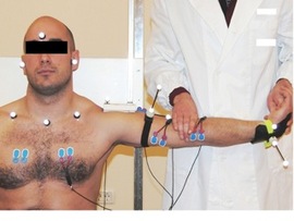
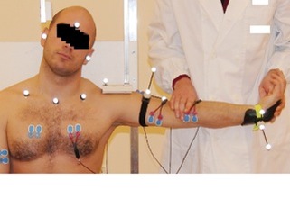
In the study by Rade et al (2012), we measured muscle function with EMG during an end range stretch as a simulation of Mills manipulation in asymptomatic subjects. The idea was to see if tension in the nerves affect the muscle response during a technique that was very similar to the clinical Mills manipulation which is normally considered a musculoskeletal technique.
One of the problems with physical techniques is specificity. This study addressed whether neural positioning could influence the effects of Mills manipulation when the neural component was changed.
ELECTROMYOGRAPHIC ACTIVITY WAS MEASURED IN THE FOLLOWING MUSCLES:
Compared with the cervical spine in neutral, we found that there was less activity in certain muscles when the cervical spine was positioned in ipsilateral lateral flexion.
Ipsilateral lateral flexion produced less activity in brachioradialis and biceps and NOT triceps, upper and lower trapezius and pectoralis major. Hence the muscle changes were in a specific pattern.
The pattern was in the muscles that protect the elbow from the movements of Mills manipulation - elbow extension and pronation. This may have occurred also because the movements of Mills manipulation (extension and pronation) also apply force to the posterior interosseous nerve and the muscles therefore have acted to protect the local nerves as well.
THE FACT THAT LESS NEURAL TENSION PRODUCED LESS MUSCULAR PROTECTION HIGHLIGHTS SOME INTERESTING POINTS:
In addition to stretching the common extensor origin, Mills manipulation probably affects the local nerves
REFERENCE
Rade M, Shacklock M, Peharec S, Bačic P, Candian C, Kankaanpää M, Airaksinen O 2012. Effect of cervical spine position on upper limb myoelectric activity during pre-manipulative stretch for Mills manipulation: A new model, relations to peripheral nerve biomechanics and specificity of Mills manipulation. Journal of Electromyography and Kinesiology 22 (2012) 363–369.
Find abstract
Purchase article
One of the problems with physical techniques is specificity. This study addressed whether neural positioning could influence the effects of Mills manipulation when the neural component was changed.
ELECTROMYOGRAPHIC ACTIVITY WAS MEASURED IN THE FOLLOWING MUSCLES:
- brachioradialis
- biceps
- triceps
- upper trapezius
- lower trapezius
- pectoralis major.
Compared with the cervical spine in neutral, we found that there was less activity in certain muscles when the cervical spine was positioned in ipsilateral lateral flexion.
Ipsilateral lateral flexion produced less activity in brachioradialis and biceps and NOT triceps, upper and lower trapezius and pectoralis major. Hence the muscle changes were in a specific pattern.
The pattern was in the muscles that protect the elbow from the movements of Mills manipulation - elbow extension and pronation. This may have occurred also because the movements of Mills manipulation (extension and pronation) also apply force to the posterior interosseous nerve and the muscles therefore have acted to protect the local nerves as well.
THE FACT THAT LESS NEURAL TENSION PRODUCED LESS MUSCULAR PROTECTION HIGHLIGHTS SOME INTERESTING POINTS:
In addition to stretching the common extensor origin, Mills manipulation probably affects the local nerves
- we may be able to increase localisation of Mills manipulation to the musculoskeletal by adding ipsilateral lateral flexion of the cervical spine which may reduce neural tissue effects at the elbow
- we may make the technique include neural tissues by adding contralateral lateral flexion. This way the nerves and musculoskeletal structures are treated simultaneously.
- as mentioned, this is a new model of investigation of possible interactions between nerve and musculoskeletal tissues which we hope will provide further insights in the future. More to come in subsequent postings.
REFERENCE
Rade M, Shacklock M, Peharec S, Bačic P, Candian C, Kankaanpää M, Airaksinen O 2012. Effect of cervical spine position on upper limb myoelectric activity during pre-manipulative stretch for Mills manipulation: A new model, relations to peripheral nerve biomechanics and specificity of Mills manipulation. Journal of Electromyography and Kinesiology 22 (2012) 363–369.
Find abstract
Purchase article